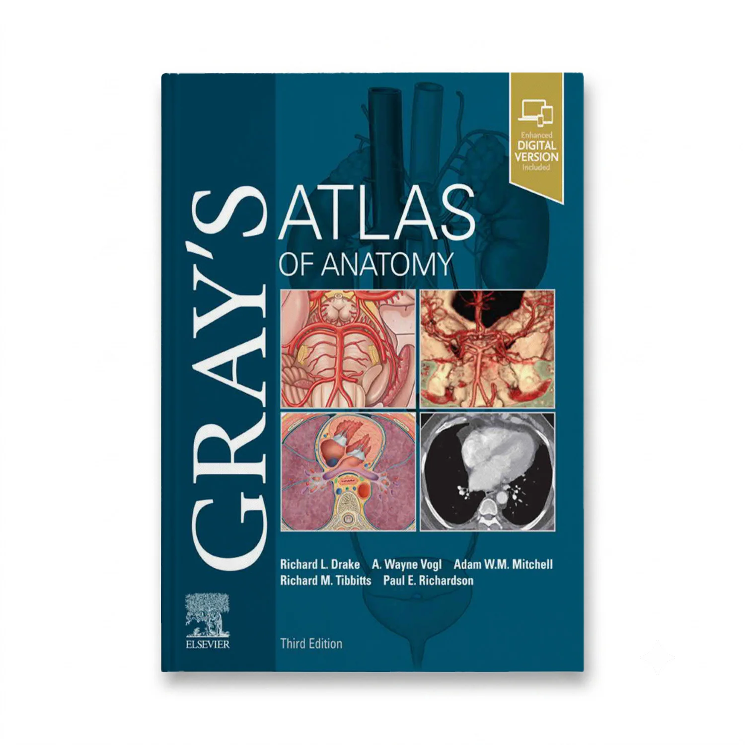
Clinically focused and consistently illustrated, Gray’s Atlas of Anatomy provides a vivid, visual depiction of anatomical structures essential for proper identification in the dissection lab and successful preparation for course exams. The companion resource to Gray’s Anatomy for Students, this edition features nearly 1,000 exquisitely illustrated anatomical figures that guide readers in identifying various body structures and understanding their relationships to one another. Surface anatomy photographs overlaid with detailed drawings help visualize the clinical correlation between anatomical structures and surface landmarks. More than 270 clinical images, including laparoscopic, radiologic, surgical, ophthalmoscopic, otoscopic, and other views, are placed adjacent to anatomic artwork for side-by-side comparison, enhancing comprehension of real-world applications. A new large-format illustration of the inguinal region in women and updated imaging figures reflect modern clinical settings. Ideal for building on existing anatomy knowledge with a logical progression from superficial to deep structures, this resource is indispensable for students and practitioners alike.

Get it before class starts
Semester is close – start reading tonight.
Right edition guaranteed
Wrong edition or ISBN? Free swap.
Help any day, any hour
File will not open? We fix it or refund you.
Buy once, keep it
No subscription, no app, no expiry date.
